
热门标签
热门文章
- 1你的服务器还安全吗?用户数据是否面临泄露风险?_连接ceye.io导致数据泄露攻击
- 2SD3开源:AI绘画的新纪元,出图效果巨好,不容错过!(附教程)_sd3 medium incl
- 3《深度思维》读书笔记_深度思维: 透过复杂直抵本质的跨越式成长方法论 pdf
- 4vivado中IP核 ila的端口数量超过64个配置方式_vivado中vio ip核设置探头数量
- 5压缩感知在图像识别中的实践
- 6LLM - 大模型评估指标之 ROUGE_evaluate.load("rouge")
- 7大模型赋能开发者!海云安荣膺毕马威中国金融科技双50榜单!
- 82024年网安最全鸿蒙ArkTS实战开发:多线程安全注意事项_ts 多线程安全(1)_ts 线程安全
- 9Selenium 与(Firefox、GeckoDriver)和(Chrome、ChromeDriver)版本对应关系_selenium4.8.3 版本对应
- 10谁可以使用.news域名?
当前位置: article > 正文
使用各向异性滤波器和图像处理方法进行脑肿瘤检测(MATLAB)
作者:人工智能uu | 2024-07-07 09:49:36
赞
踩
使用各向异性滤波器和图像处理方法进行脑肿瘤检测(MATLAB)
医学图像分割一直以来都是计算机辅助诊断领域的研究热点。在医学图像的处理和分析中,对图像中感兴趣区域的准确分割尤其关键。要对感兴趣区域进行分类识别,首先要从图像中把感兴趣区域精确分割出来,然后有针对性地对感兴趣区域提取特征并分类。目前关于脑部MRI的分割主要的研究都集中在脑部的灰质,白质和脑脊液等组织的分割。
图像分割方法根据分割类型的不同,大致可以分为基于区域的分割方法和基于边界的分割方法。基于区域的分割方法一般是根据图像中点与点之间的灰度值、纹理特征等属性来判断它们的相似度,根据相似度来划分图像区域。基于边界的分割方法是以灰度变化的梯度大小来确定感兴趣区域的边界。在实际应用中,常常将这两种方法结合在一起使用。
鉴于此,采用各向异性滤波器和图像处理方法进行脑肿瘤分割检测,运行环境为MATLAB 2018,主运行代码如下:
-
-
- %%filter
- num_iter = 10;
- delta_t = 1/7;
- kappa = 15;
- option = 2;
- disp('Preprocessing image please wait . . .');
- inp = anisodiff(s,num_iter,delta_t,kappa,option);
- inp = uint8(inp);
-
- inp=imresize(inp,[256,256]);
- if size(inp,3)>1
- inp=rgb2gray(inp);
- end
- figure;
- imshow(inp);
- title('Filtered image','FontSize',20);
-
- %%thresholding
- sout=imresize(inp,[256,256]);
- t0=60;
- th=t0+((max(inp(:))+min(inp(:)))./2);
- for i=1:1:size(inp,1)
- for j=1:1:size(inp,2)
- if inp(i,j)>th
- sout(i,j)=1;
- else
- sout(i,j)=0;
- end
- end
- end
-
- %morphologial operation
- label=bwlabel(sout);
- stats=regionprops(logical(sout),'Solidity','Area','BoundingBox');
- density=[stats.Solidity];
- area=[stats.Area];
- high_dense_area=density>0.6;
- max_area=max(area(high_dense_area));
- tumor_label=find(area==max_area);
- tumor=ismember(label,tumor_label);
- if max_area>100
- figure;
- imshow(tumor)
- title('tumor alone','FontSize',20);
- else
- h = msgbox('No Tumor!!','status');
- %disp('no tumor');
- return;
- end
-
- %%binding box
- box = stats(tumor_label);
- wantedBox = box.BoundingBox;
- figure
- imshow(inp);
- title('Bounding Box','FontSize',20);
- hold on;
- rectangle('Position',wantedBox,'EdgeColor','y');
- hold off;
- %%getting tumor outline
- dilationAmount = 5;
- rad = floor(dilationAmount);
- [r,c] = size(tumor);
- filledImage = imfill(tumor, 'holes');
- for i=1:r
- for j=1:c
- x1=i-rad;
- x2=i+rad;
- y1=j-rad;
- y2=j+rad;
- if x1<1
- x1=1;
- end
- if x2>r
- x2=r;
- end
- if y1<1
- y1=1;
- end
- if y2>c
- y2=c;
- end
- erodedImage(i,j) = min(min(filledImage(x1:x2,y1:y2)));
- end
- end
- figure
- imshow(erodedImage);
- title('eroded image','FontSize',20);
-
- %%subtracting eroded image from bw image
- tumorOutline=tumor;
- tumorOutline(erodedImage)=0;
- figure;
- imshow(tumorOutline);
- title('Tumor Outline','FontSize',20);
-
- %%inserting outline
- rgb = inp(:,:,[1 1 1]);
- red = rgb(:,:,1);
- red(tumorOutline)=255;
- green = rgb(:,:,2);
- green(tumorOutline)=0;
- blue = rgb(:,:,3);
- blue(tumorOutline)=0;
- tumorOutlineInserted(:,:,1) = red;
- tumorOutlineInserted(:,:,2) = green;
- tumorOutlineInserted(:,:,3) = blue;
- figure
- imshow(tumorOutlineInserted);
- title('Detected Tumer','FontSize',20);
-
-
- %%to display
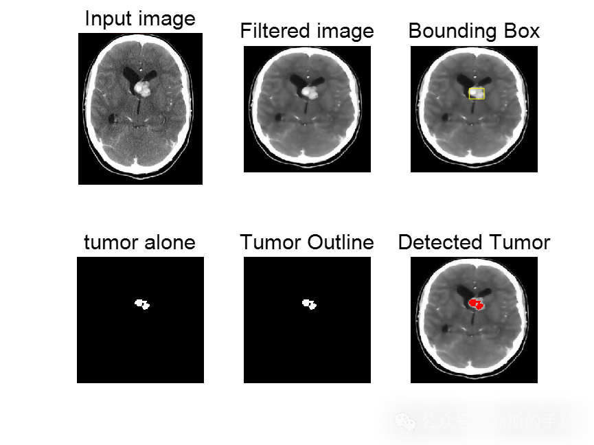
- figure
- subplot(231);imshow(s);
- title('Input image','FontSize',15);
- subplot(232);imshow(inp);
- title('Filtered image','FontSize',15);
- subplot(233);imshow(inp);
- title('Bounding Box','FontSize',15);
- hold on;
- rectangle('Position',wantedBox,'EdgeColor','y');
- hold off;
- subplot(234);
- imshow(tumor);
- title('tumor alone','FontSize',15);
- subplot(235);imshow(tumorOutline);
- title('Tumor Outline','FontSize',15);
- subplot(236);imshow(tumorOutlineInserted);
- title('Detected Tumor','FontSize',15);
-
- 知乎学术咨询:
- https://www.zhihu.com/consult/people/792359672131756032?isMe=1
- 工学博士,担任《Mechanical System and Signal Processing》《中国电机工程学报》《控制与决策》等期刊审稿专家,擅长领域:现代信号处理,机器学习,深度学习,数字孪生,时间序列分析,设备缺陷检测、设备异常检测、设备智能故障诊断与健康管理PHM等。
-


- 工学博士,担任《Mechanical System and Signal Processing》《中国电机工程学报》《控制与决策》等期刊审稿专家,擅长领域:现代信号处理,机器学习,深度学习,数字孪生,时间序列分析,设备缺陷检测、设备异常检测、设备智能故障诊断与健康管理PHM等。
声明:本文内容由网友自发贡献,不代表【wpsshop博客】立场,版权归原作者所有,本站不承担相应法律责任。如您发现有侵权的内容,请联系我们。转载请注明出处:https://www.wpsshop.cn/w/人工智能uu/article/detail/795309
推荐阅读

相关标签

